In our Featured Case series, NuVasive highlights outcome driven innovation, and the combination of access, interbody implants, biologics, fixation and enabling technologies to create a comprehensive system to address patient pathologies.
In this case, Dr. Chase Bennett, Spine Surgeon, Novant Health Brain and Spine Surgery—Kimel Park, performs a multi-staged thoracic pedicle subtraction osteotomy and spinal fusion using Coroent Small Interlock, Vuepoint 2, Reline, Bendini OCT and NuVasive Power to correct hyper kyphosis and restore lordosis in a patient suffering from ankylosing spondylitis.
“Cervicothoracic reconstruction is not for everybody—and the reason you’d need it is not very common, but it can offer a great deal of pain relief and improvement in quality of life for the right person.”
– Dr. Chase Bennett
Case details:
- Gender: Male
- Age: 64
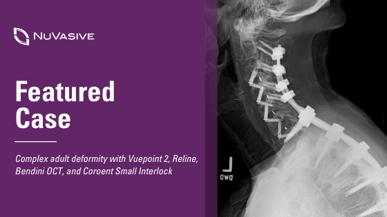
- History: Neck pain with secondary back pain
- Diagnosis: Ankylosing spondylitis resulting in thoracic hyper kyphosis; loss of cervical lordosis
Surgical plan:
Dr. Bennett determined that the best course of action to correct the patient’s hyper kyphosis and restore lordosis in the cervical and upper thoracic spine would involve a complex two-stage procedure. The first stage of the procedure involved a C3-C7 ACDF utilizing CoRoent Small Interlock interbody implants to restore sagittal alignment in the cervical spine. This procedure was followed by the second stage which involved a pedicle subtraction osteotomy (PSO) at T1 and posterior fixation from C2-T5 utilizing Bendini, Vuepoint 2, navigation and Reline 3CO.
- Procedure:
- Stage 1: C3-C7 ACDF
- Stage 2: T1 PSO; C2-T5 posterior fixation
- Interbody: Coroent Small Interlock
- Fixation: Vuepoint 2, Reline
- Instrumentation: Reline 3CO rack, NuVasive Power
- Enabling technologies: Intraoperative navigation; Bendini OCT
The ACDF was completed on the first day followed by the remaining steps of the procedure on the second. In the second stage of the procedure, his partner, Dr. Ross McEntarfer, Spine Surgeon, Novant Health Brain and Spine Surgery—Kimel Park, who has additional expertise with PSO joined Dr. Bennett for this more complex stage of the surgery.
“Having an assistant or co-surgeon who understands the procedure and can provide a second set of skilled hands was essential for patient safety and to move the procedure forward in a timely manner. A senior resident, fellow or partner may fulfill this role. Excellent communication and intraoperative collaboration benefits all parties involved. ”
– Dr. Chase Bennett
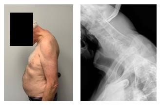
In the second stage of the procedure, Drs. Bennett and McEntarfer did laminectomies at C7 and T1 and hemilaminectomies at C6 and T2 to allow for visualization of the spinal cord. The team then proceeded with the T1 PSO utilizing the Reline 3CO rack and closely monitoring motors with NVM5, allowing them to move efficiently through the steps and bilaterally close the osteotomy to restore sagittal alignment. Bendini was utilized for bending the rod. This time saving technology facilitated a precise bend in the rod allowing the rack compressors to close the osteotomy and minimized the need for additional reduction.
Intraoperative navigation and NuVasive Power were a benefit for this procedure in multiple ways. When completing the PSO, navigated instruments facilitated an accurate anterior depth for bony removal. Navigating pedicle screws at C7 and in the upper thoracic spine allowed for excellent fixation across the osteotomy site and utilizing power effectively balanced speed and maneuverability. Many PSOs will utilize a quad-rod technique across an osteotomy site. With a smaller Reline pedicle screw utilized at C7, a transition rod was placed to allow the 5.5 mm diameter rod to cross the osteotomy and eliminate the need for supplemental hardware.
“Several key technologies were used in this case. I placed my C7 and thoracic pedicle screws under power and with navigation. The use of power minimizes deflection and maximizes navigation accuracy by minimizing force applied to the spine. Additionally, the drill rotates completely coaxially with the screw and minimizes off axis precession. Navigation is also very useful for guiding the depth and extent of the osteotomy as well as any necessary rib resections. Finally, there are several great tools in the 3CO set. In particular, I feel the posterior wall pusher is more effective than using a tamp or down-going curette, and using the rack-compressor across the C7 and T2 screws enables local angulation across the osteotomy when manipulating the Mayfield.”
– Dr. Chase Bennett
Outcome:
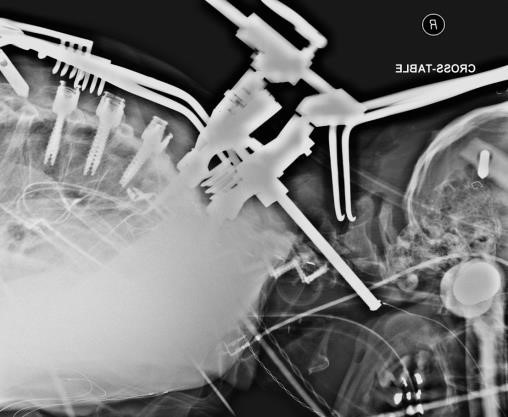
Dr. Bennett was able to restore a normal gaze and cervical lordosis for the patient through this procedure. The patient was on his feet the next day and began physical therapy within nine days of his procedure. Five months into his recovery, he is at or ahead of schedule, expecting to return to normal activity within a year.
Learn more about the patient’s journey and the procedure at Novant Health.
An individual’s surgical procedure and recovery may deviate from what is described herein. Each case is unique and has its own independent considerations. It is the surgeon’s responsibility to discuss all relevant risks with the patient prior to surgery.
Click here to access each of the above-referenced product specific Instructions for Use for important product safety information, including, but not limited to, indications, contraindications, warnings, precautions, and potential adverse effects.
Click here to read more featured cases.
9513556 A