Attrax®

Attrax is a unique ceramic bone graft substitute with an optimized surface microarchitecture designed to drive bone fusion. Attrax Putty is the first and only ceramic supported by a Level I randomized controlled trial as a bone graft substitute in posterolateral lumbar fusions (PLF).1

The proof is in the putty.

Level I clinical evidence

A multicenter, randomized, intra-patient, controlled trial investigated the efficacy of Attrax Putty as a bone graft substitute for autograft in instrumented PLF. For each patient, Attrax Putty was randomized to one side of the posterolateral spine and autograft (at least 50% iliac crest bone graft) was placed on the opposite side. The major advantage of this study design was the elimination of interpatient variability. Fusion was assessed at one year follow up on CT-scans, with each level and each side scored by blinded observers. The study concluded Attrax Putty alone successfully demonstrated non-inferiority compared to autograft in instrumented posterolateral spinal fusions.2 

Preclinical fusion performance

Fusion superior to traditional synthetic ceramic grafts

Spinal segments fused with Attrax had greater biochemical strength than segments treated with Actifuse ABX or Vitoss® BA in a rabbit PLF model.3

Statistically lower than Attrax Putty (P<0.05).

Fusion equivalent to autograft

In a large animal instrumented posterolateral fusion (PLF) model, Attrax fusion rates were equivalent to or better than autograft, and faster than traditional synthetic.4-6*

*Data from intramuscular and spine preclinical models may not be representative of clinical outcomes.

Optimized microarchitecture

Deliberately engineered. Intelligently designed.

The Attrax ceramic surface has unique microstructure and microporosity that are optimized for bone formation. The unique microarchitecture of Attrax drives the differentiation of mesenchymal stem cells (MSCs) into bone-forming osteoblasts without added growth factors.7

The optimized microarchitecture of Attrax is engineered using tightly controlled parameters for a defined micropore size distribution within 0.3 to 1.1 microns. Traditional calcium phosphate materials falling outside of this specification do not possess the unique ability of Attrax to form bone consistently intramuscular defects.

BIO-2292_AttraxMicrositeUpdates-images_Key
BIO-2292_AttraxMicrositeUpdates-images_PoreConcentrationG
AttraxMicrosite_UpdatedImages_Attrax
AttraxMicrosite_UpdatedImages_Nanostructured
AttraxMicrosite_UpdatedImages_Traditional

Learn more about Attrax

Interested in more information or trying a free sample?

Get in touch with our Biologics team

Learn more about Attrax

Interested in more information or trying a free sample?

Get in touch with our Biologics team

1. Based on review of publicly available materials at the time of this release.
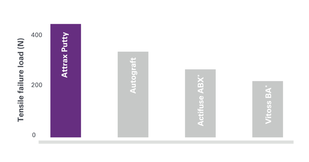
2. Lehr AM, Oner FC, Delawi D, et al. Efficacy of a standalone microporous ceramic versus autograft in instrumented posterolateral spinal fusion. Spine 2020;45(14):944-51.
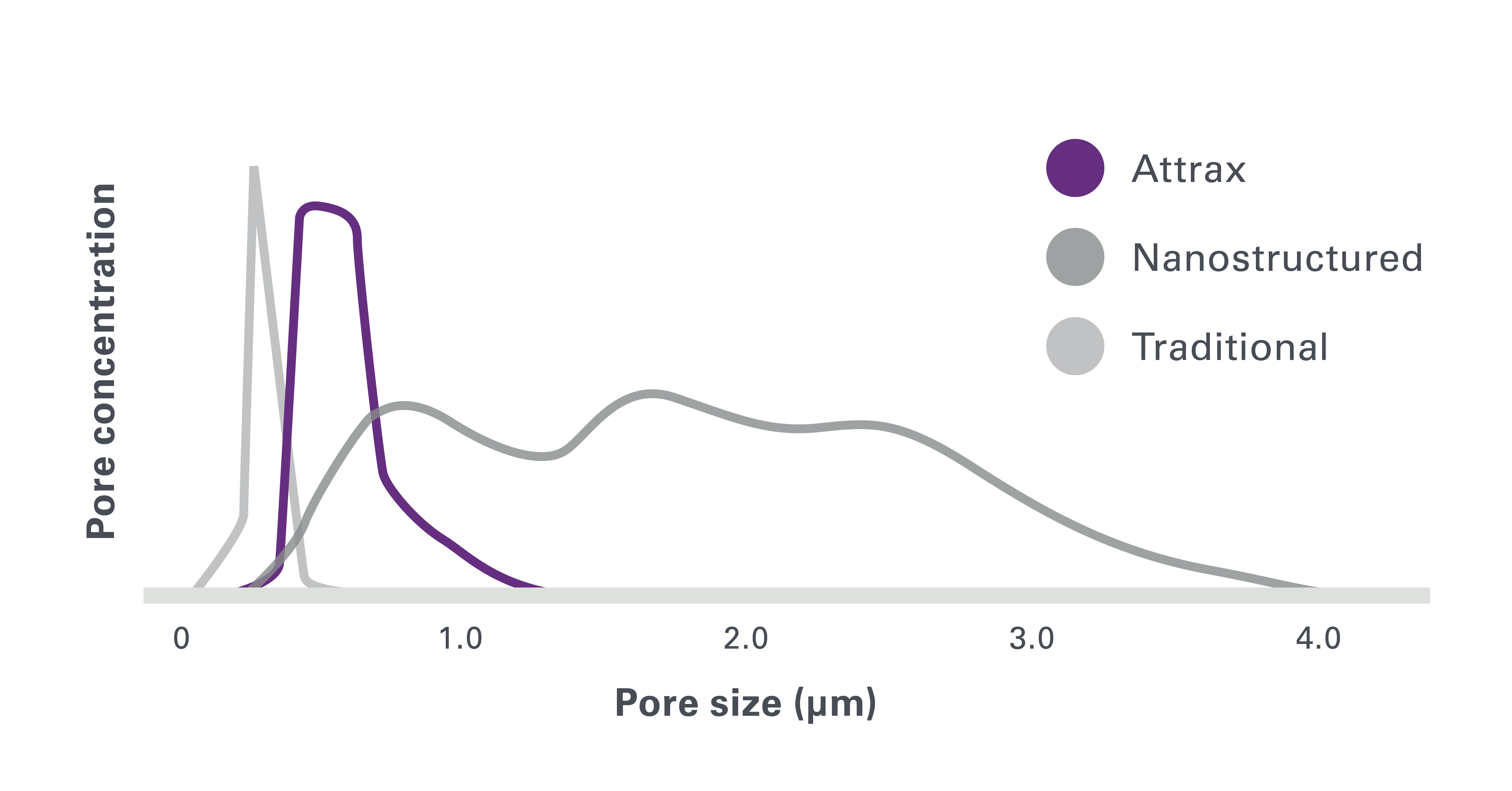
3. Walsh WR, Degroot F, Bertollo N, et al. Nanostructured TCP in rabbit posterolateral fusion compared to commercial osteobiologics. American Academy of Orthopaedic Surgeons 2011 Annual Meeting. San Diego, CA, USA.
4. Ismailoglu AS, Vizesi F, Cunningham B, et al. Fibrillar collagen/ TCP scaffold in the sheep posterolateral fusion model. Society for Biomaterials Annual Meeting; 2012; New Orleans, LA, USA.
5. Fredericks DC, Smucker JD, Peterson EB, et al. Novel TCP compares favorably to autograft in posterolateral fusion: evaluation in rabbit and sheep models. International Society for the Advancement of Spine Surgery Annual Conference; 2013; Vancouver, BC, Canada.
6. Barbieri D, Yuan H, Ismailoglu AS, et al. Comparison of two moldable calcium phosphate-based bone graft materials in a noninstrumented canine interspinous implantation model. Tissue Eng Part A 2017;23(23- 24):1310-20.
7. Yuan H, Fernandes H, Habibovic P, et al. Osteoinductive ceramics as a synthetic alternative to autologous bone grafting. PNAS 2010;107(31):13614-9.